
Low Vision
– by Dr. Shaher Saad Eddin
Definition of low vision
When the patient’s best correction is applied (e.g., glasses), with both eyes and the following constraints or difficulties are present. In simple words: Not eligible to drive and may have difficulties recognizing faces across a street, watching television, or choosing clean, unstained, coordinated clothing.
| A visual acuity of less than 6/18 but greater than 3/60. |
| Not eligible to drive and may have difficulties recognizing faces across a street, watching television, or choosing clean, unstained, coordinated clothing. |
| The patients’ visual field 10° or less. |
| or the contrast sensitivity is less than 5%. |
There are congenital or acquired reason for Low Vision or Visual Impariment
This severely visually impaired result of ocular disorders and diseases either congenital or acquired which requires a Low Vision Aid. There are pathological diseases, disorders and dystrophies which cause a severe drop of vision leading to “visual impairment” such as: (WHO list of some explained in details)
| Diabetic retinopathy |
| Age related macular degeneration / AMD |
| Glaucoma |
| Retinitis pigmentosa – RP |
| Retinopathy of prematurity – ROP |
| Stargardt’s disease |
| Albinos- Oculocutaneous albinism |
| Rod-Cone Dystrophy |
The visual assessments for low vision patients:
The patient’s medical history (Anamnesis) such as medication or general physical health, injuries etc..
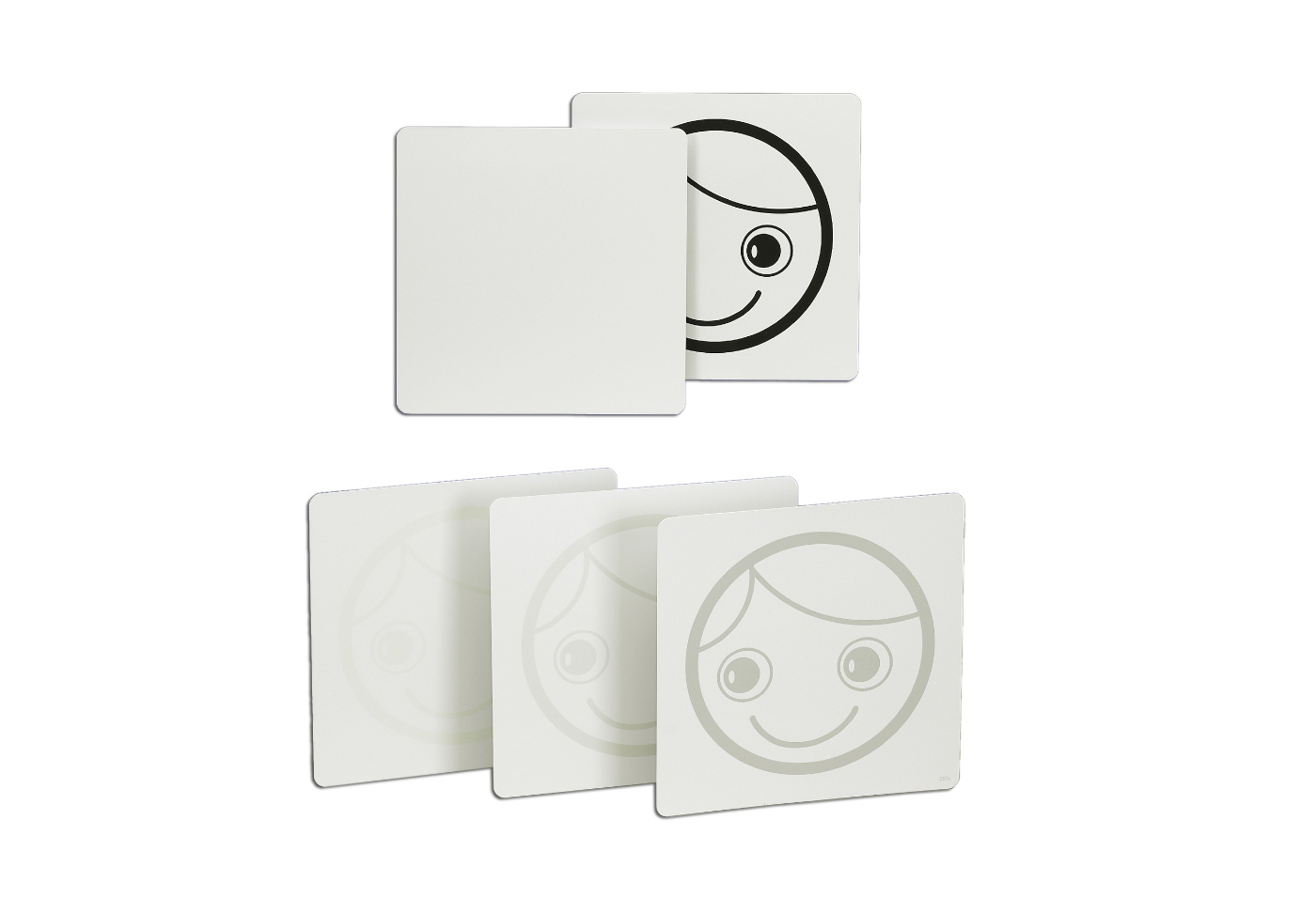
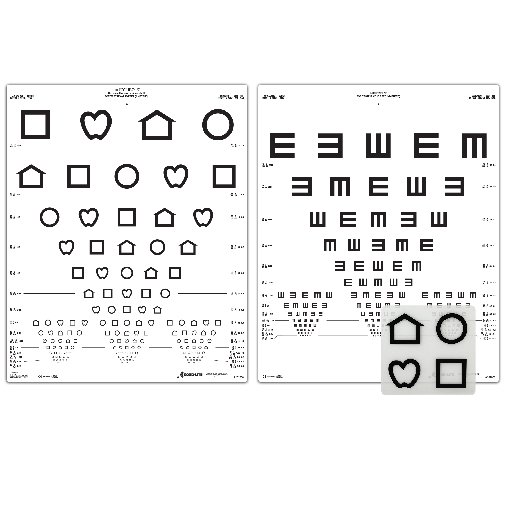
The visual acuity tests by using special charts such as „Lea Charts“ for a distance of 3m and at the near range of 40 cm. The first version of the LEA test was developed in 1976 by Finnish pediatric ophthalmologist Lea Hyvärinen, MD, PhD.
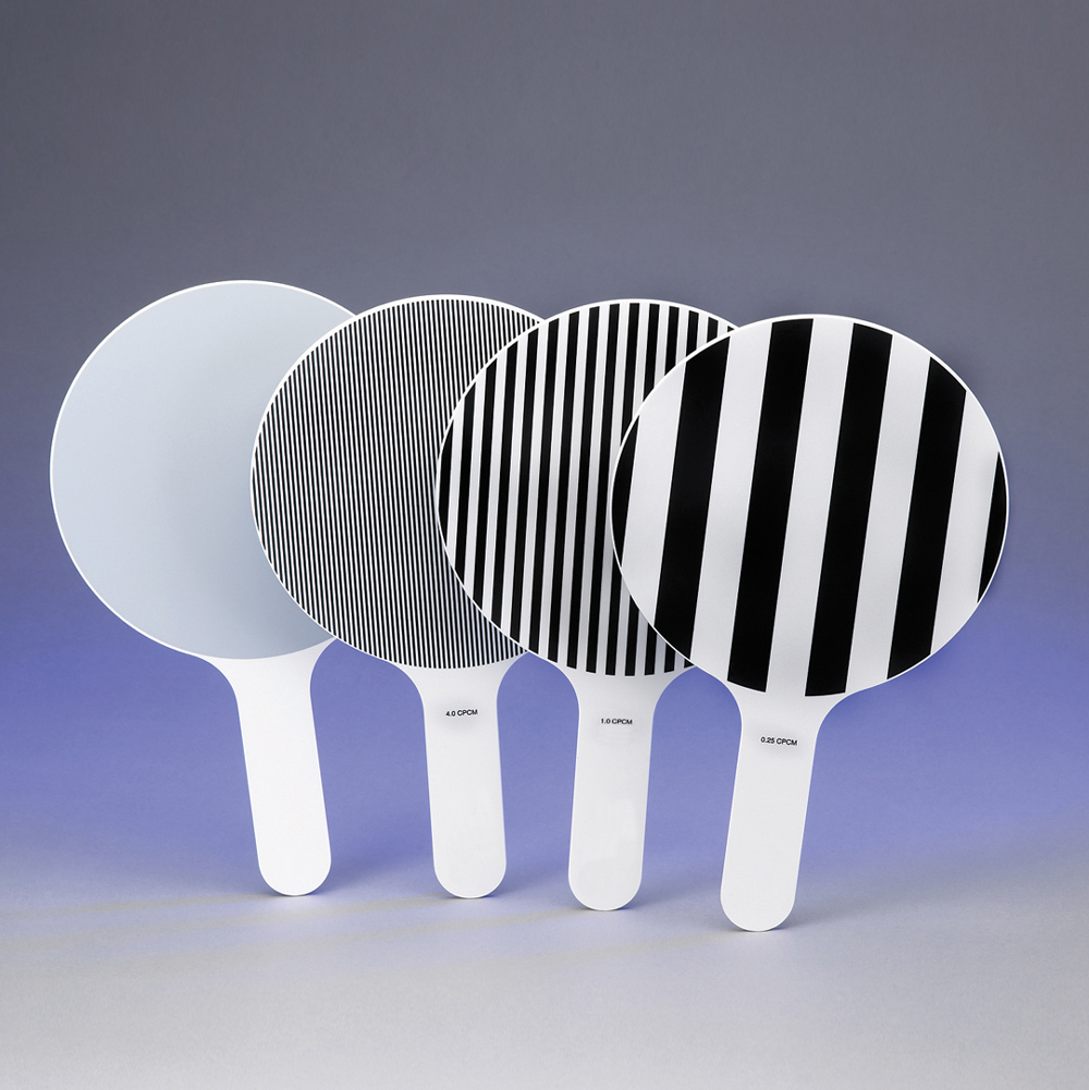
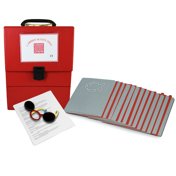
Furthermore there are the Cardiff test or Haag Streit test, or contrast sensitivity test by using special charts:
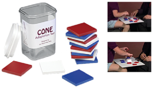
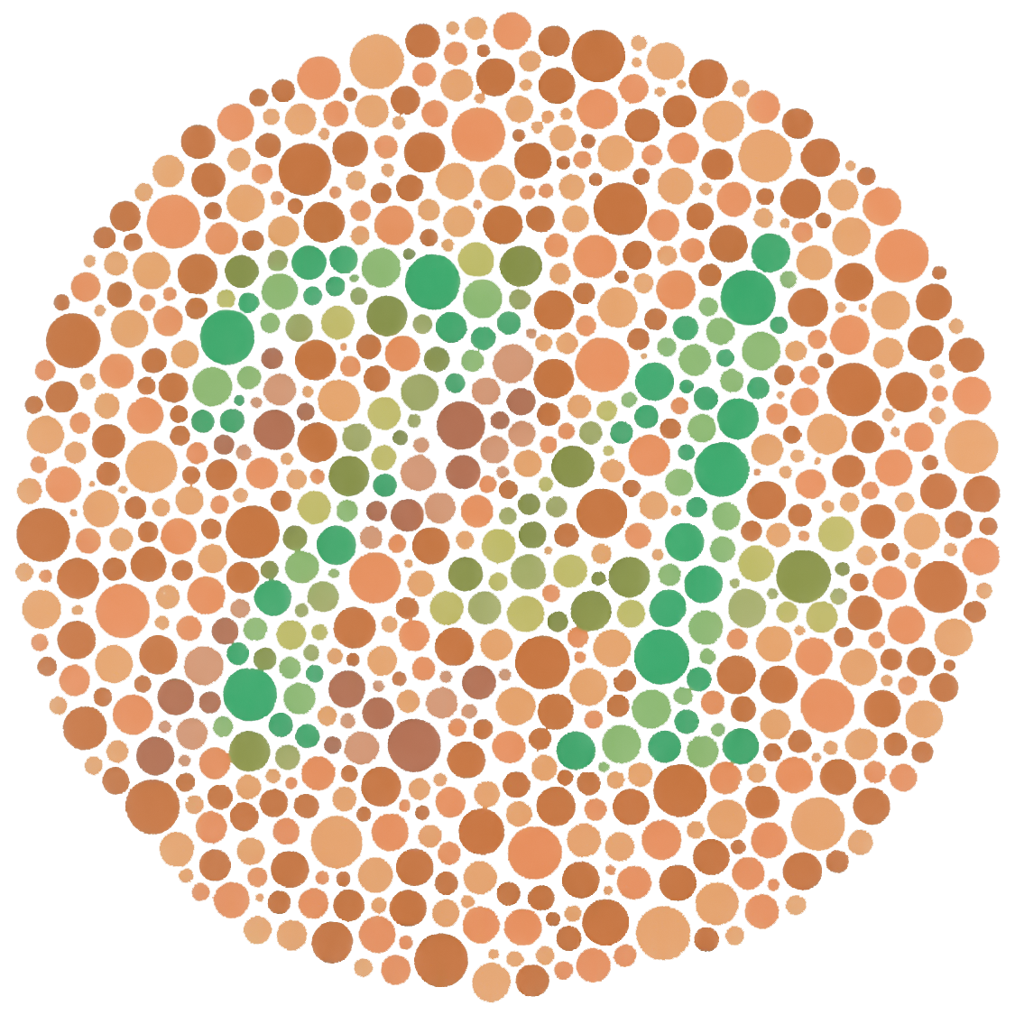
Such as eye-hand-coordination or P.O. BOX Test, cone adaptation test to diagnose scotopic and photopic functions, Color Colum bar test for color deficiency and cone adaptation, Ishihara test for color deficiency
Visual Assessment
Refraction: [determination of (eyeglass) lens prescription]. An uncorrected refractive error is a significant cause of reduced visual acuity, even in the presence of ocular disease or a visual disorder.
When the refractive error of the patient is corrected, it will affect the success of low vision aid devices. The evaluation of the refractive status, so called objective (cooperation of the patient is not necessary) and subjective (patient answers question to compare visual impressions) tests are used to determine the new prescription as a prerequisite for the further assessment of the low vision devices.
Objective and subjective refractive error test:
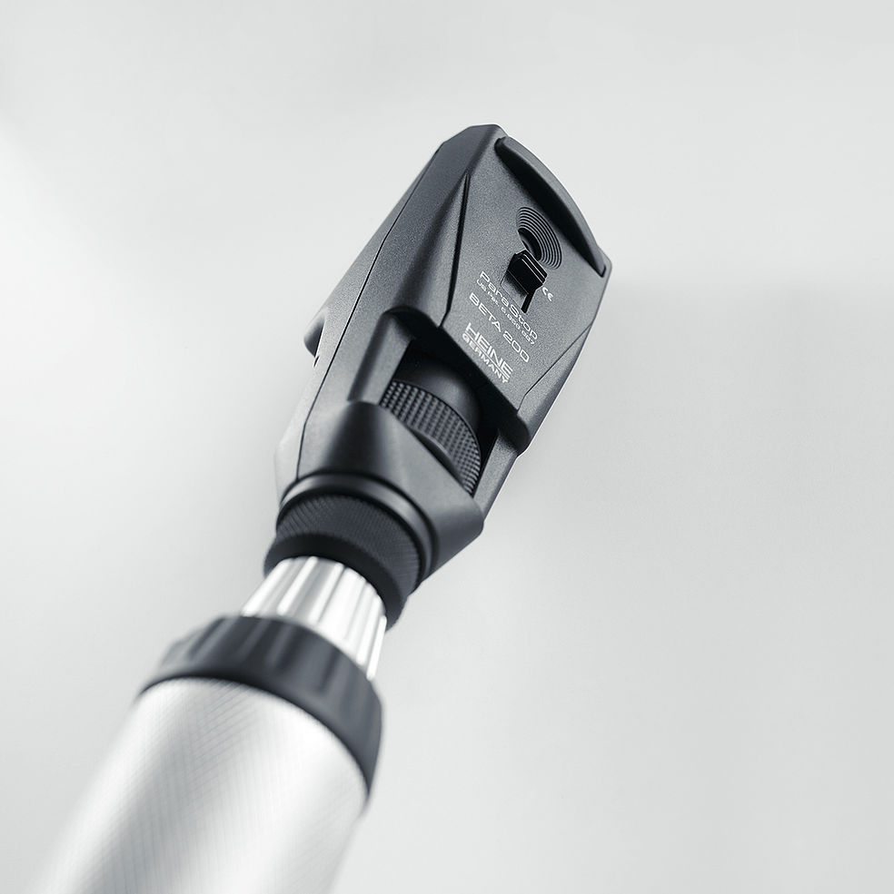
| Retinoscope: The most versatile device to evaluate the refractive error objective. Especially when media opacities, small pupils are present. This is an objective refraction test. |
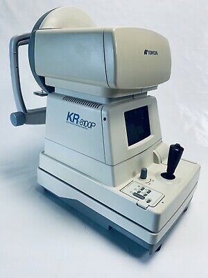
| Autorefractometer: Very common computerized device with electronic, mechanical automatic objective measurement. This is an objective refraction test. |
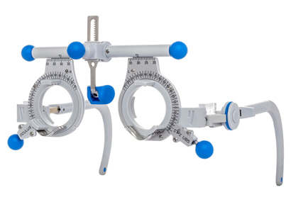
| Trial frame or phoropter for the subjective refractive error test. This is an subjective refraction test. |
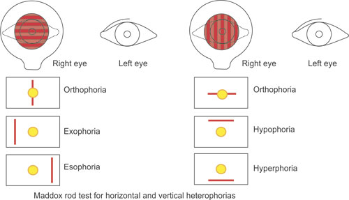
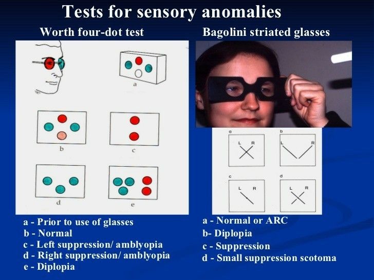
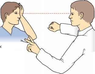
Ocular motility and binocular vision tests
The oculomotor system must be evaluated for the presence of nystagmus, ocular motility dysfunction (poor saccades or pursuits), strabismus and diplopia.
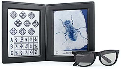
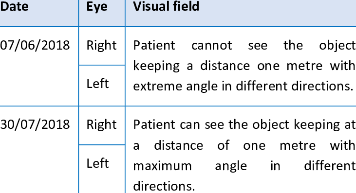
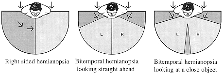
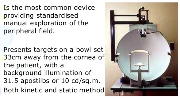
Visual Field Assessment
There are international tests and devices specialized to diagnose, investigate and treat all low vision patients with different ages, different ocular diseases, disorders and dystrophies.
Low vision aid devices
There are low vision aids devices available to help the patient not only to improve the vision but also to improve the quality of life for oneself, the family and to achieve the visual requirements and personal goals.
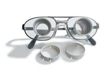
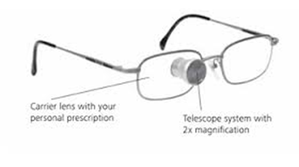
The telescopic system (telescopic glasses), Galileo Galilei binocular (two eyes) or monocular for one eye only to improve distance and intermediate range.
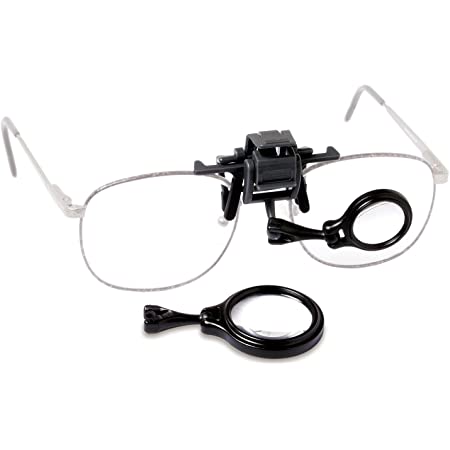
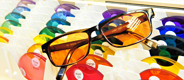
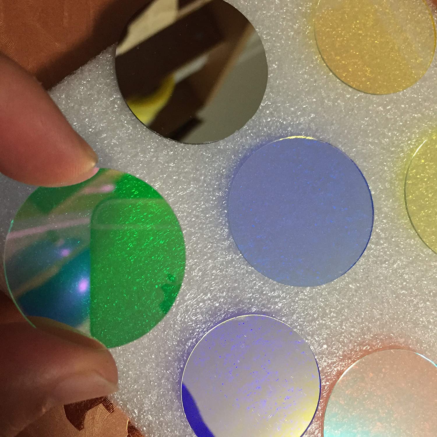
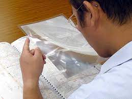
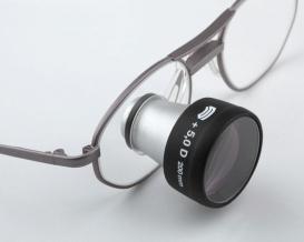
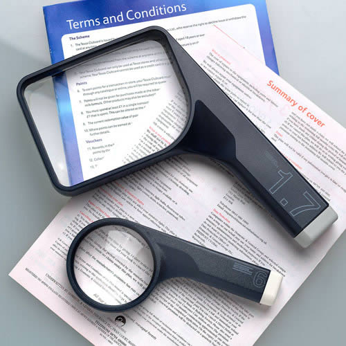
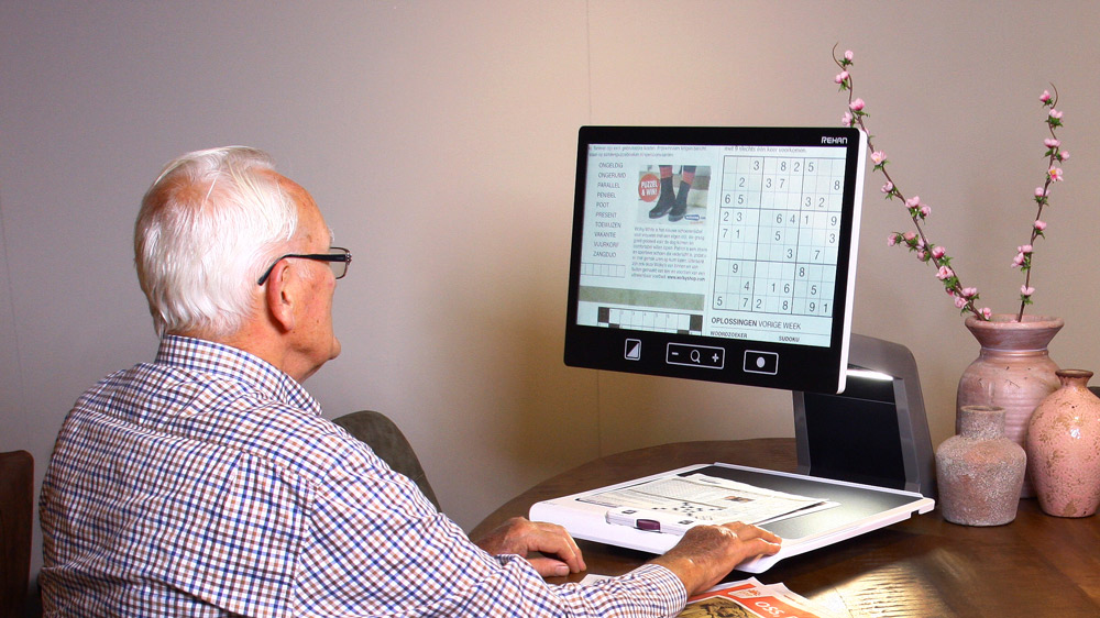
The Kepler monocular is mainly used for higher magnifications at the near vision range. Magnifiers for reading vision like clip-on and hands magnifiers. To improve near vision and reading ability. Ophthalmic Filters lenses for contrast sensitivity enhancement and to reduce photophobia and Light/Dark adaptation time, Clip-On or fixed lenses. Prismatic glasses for near and distance vision – to increase the visual field in some cases of Glaucoma and Nystagmus, or treating the strabismus with Fresnel prism or fixed prismatic lenses. Electronic magnifier-for near vision
Non-optical aids, Computer software and Computer Reading Screen Reading devices
Cases:
- Patient with diabetic retinopathy the best correct vision with both eye was 6/60 with low vision aid telescopic magnifier 2.1x become V.A 6/24
- Patient with RP (Retinitis Pigmentosa) the vision at day light was very impaired 6/48, with outdoor filter the vision become 6/12.
- AMD patient with a near vision, M5 with prismatic reading glasses become M3 and with electronic magnifier become M1.
Feel free to drop any question in the commentary section. Best wishes.
The mission is the vision.


