
The preparation for any surgery or any medical treatment makes an anamnesis mandatory and is a routine exercised by every medical staff. The refractive surgery performed on a human eye to change the vision when the patient wishes not to use glasses or contact lenses is quite common. The standard investigation regime to determine the status of the eye to receive the surgery consists of some mandatory test results already. The following should have an additional focus on before performing the refractive surgery

Refractive Error and Refractive Power – Eye Examination
Starting with the refractive error test, the surgeon must do a full investigation. The visual acuity evaluated when the ‘old’ glasses or contact lenses are added and without any correction is telling us helpful information regarding the patient’s ‘visual behavior’ and is a mandatory procedure. A full refractive error test via a retinoscopy follows the standard investigation, followed by a cycloplegic refraction.
Prerequisite Facts
The prescription of the corrective power for glasses or contact lenses, also called the ‘refraction’, must be stable for more than 6 months before the refractive surgery. What power of the refractive error can we tackle with a refractive surgery? For a myopia (shortsightedness) up to-10 Diopter and a hyperopia (farsightedness) up to +6.00 Diopter, astigmatism up to 6.00Diopter.
Important Steps
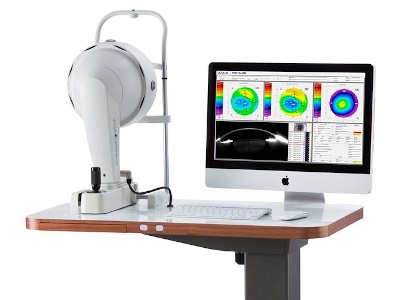
The things we focus at next are the corneal topography. At the eye specialty hospital in Jordan, Amman, we use the OCULAS-PENTACAM.
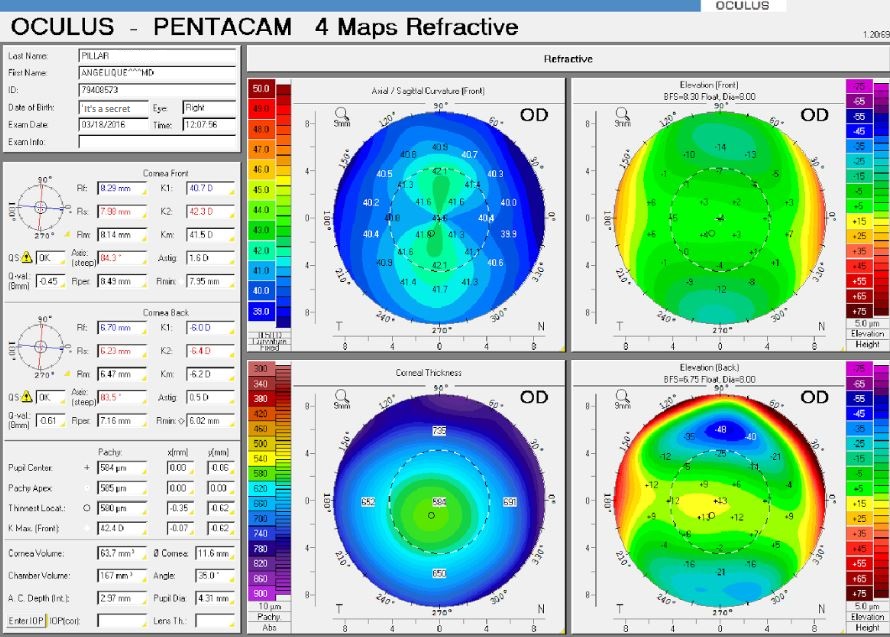
We treat the refractive error by changing the corneal curvature, so the cornea should be clear and healthy, to investigate if this situation is present, we use the pentacam to generate a graphical picture of the corneas surface in conjunction with all the geometrical data via a very advanced system.
There are specific details to focus on the pentacam is presenting us before we go ahead and do the refractive surgery.
First
The sagittal curvature which gives us the corneal curvature perimeters called K1, K2 and the average Km (K is D = Diopter). The Km after the operation should not be less than 36D and not more than 48D. For each (-) Diopter of the refractive error we apply a decrease of 0.8 curvature Diopter, and for each (+) Diopter we increase 0.8 curvature Diopter.
For example, if we want to treat a refractive error of -2.00 sphere Diopter and the Km = 44D, after treatment the Km will be 42.4D. And if we want to treat +2.00 sphere Diopter vice versa the Km will be 45.6D after the treatment.
Second
we must focus on the thinnest location of the corneal thickness. For this we have a special nomogram, an image-generating procedure which measures the thickness of the cornea before and after the operation. The general rule of the application is for each 1 Diopter the thickness of the cornea will decrease 15nm due to the operation.
For example, if the refractive error is -2.00 sphere Diopter and the corneal thickness at the thinnest location was before the operation 500nm, after the operation it will be 476nm.
There are deferent nomograms for each operation, e.g. PRK and Lasik. For the PRK the corneal thickness should not be less than 400nm and for Lasik not less than 300nm.
Third
The pupil diameter must not be more than 5 mm at normal light conditions because if we treat a patient, e.g. 5.50 mm pupil diameter, there is a likelihood, to suffer of night glare after the operation.
Corneal Hysteresis
At the eye specialty hospital, we use a device to determine the corneal hysteresis, which gives us an important insight and diagnosis regarding the rigidity of the corneal tissue and the ability to absorb and dissipate energy. We use this device to select the treatment procedure for the patient. E.g., if the corneal hysteresis (CH) is less than 7, we do not perform the operation and if it is between 7.5 to 8.5 we can do the PRK and if it is more than 8.5 we can go for a LASIK operation.
Dry Eye
The investigation into eye dryness. In case, the patient suffers of a moderate or even to a severe eye dryness we do not perform the operation.
Finally
The ophthalmology surgeon must do a slit lamp examination of the eye with all the holistic aspects before the refractive surgery. This includes, just to mention the most important one, the eye sections such as the examination of the anterior and posterior chamber including the angles and sides of the eye chambers.
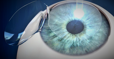
The retina, which is of special focus for a LASIK treatment due to the exposure of the eyeball of a high pressure through the suction when the corneal flap is produced.
Greetings from Dr. Shaher


